Home
/ Leg Bones Diagram - Bones Healing Healthy Holistic : Numerous bone is the long bone of the upper arm which goes all the way to the elbow.
Leg Bones Diagram - Bones Healing Healthy Holistic : Numerous bone is the long bone of the upper arm which goes all the way to the elbow.
Leg Bones Diagram - Bones Healing Healthy Holistic : Numerous bone is the long bone of the upper arm which goes all the way to the elbow.. Dog leg bone diagram / dog anatomy leg bones stock image stock photo download image now istock / paw bone between the heel and the phalanges.license image the bones of the leg are the femur, tibia, fibula and the foot bones shown in this diagram are the talus, navicular, cuneiform, cuboid, metatarsals and from dogs with three legs to cats without eyes, the perfect imperfection photo series. The bones of the leg are the femur, tibia, fibula and patella. Some types of leg pain can be traced to problems in your lower spine. Long bone femur label 12 photos of the long bone femur label , bone. The thigh bone, or femur, is the large upper leg bone that connects the lower leg bones (knee joint) to the pelvic bone (hip joint).
The foot bones shown in this diagram are the talus, navicular, cuneiform, cuboid, metatarsals and calcaneus. Most leg pain results from wear and tear, overuse, or injuries in joints or bones or in muscles, ligaments, tendons or other soft tissues. These muscles work together to produce movements such as standing, walking, running, and jumping. The pubis, ischium, and ilium together constitute the pelvis while the thigh bone is the femur. Hip and leg bone diagram / lower leg bones anatomy anatomy drawing diagram / this lengthy bone connects with the knee at one finish and the ankle on the different.
Foot Anatomy Bones The Talus Bone Supports The Leg Bones Tibia And from hotel-prachtig.fun Long bone femur label 12 photos of the long bone femur label , bone. Health diagram bone skeleton leg knee science anchor chart human human body. The tibia and the fibula, at the top of the ankle joint. The tibia and fibula are two long bones that run parallel to each other forming the scaffold of the leg and providing attachment points for many muscles. The foot bones shown in this diagram are the talus, navicular, cuneiform, cuboid, metatarsals and calcaneus. The tibia and fibula are two long bones that run parallel to each other, forming the scaffold of the leg and providing attachment points for many muscles. Leg bones diagram diagram schematic ideas lower leg muscle diagram blank sketch coloring page antique 1890s medical anatomy diagram leg bones skeleton posted on april 18, 2019april 18, 2019. Premier leg skeleton with painted & labeled muscle attachments.
The tibia and fibula are two long bones that run parallel to each other forming the scaffold of the leg and providing attachment points for many muscles.
Related posts of diagram of leg bones long bone femur label. Its lower end helps create the knee joint. The lower leg is comprised of two bones, the tibia and the smaller fibula. Ankle bones anatomy, arm bones anatomy, fibula anatomy, fibula fracture, hip bones anatomy, leg bones human body, foot, ankle bones anatomy, arm bones anatomy, fibula anatomy, fibula fracture, hip bones anatomy, leg bones human body. The bones of the leg are the femur, tibia, fibula and patella. The knee joint is the largest joint in the body and is primarily a hinge joint, although some sliding and rotation occur. Premier leg skeleton with painted & labeled muscle attachments. The bones of the leg and foot form part of the appendicular skeleton that supports the many muscles of the lower limbs. The femur, or thigh bone, is the single bone of the thigh region (figure 6.51). The forearm is the long bone that runs just after the elbow. The thigh bone, or femur, is the large upper leg bone that connects the lower leg bones (knee joint) to the pelvic bone (hip joint). Posted on june 4, 2014 by admin. 15 photos of the leg bones anatomy diagram.
The pubis, ischium, and ilium together constitute the pelvis while the thigh bone is the femur. The tibia and the fibula, at the top of the ankle joint. This diagram depicts diagram leg bones anatomy. Related posts of diagram of leg bones long bone femur label. The foot bones shown in this diagram are the talus, navicular, cuneiform, cuboid, metatarsals and calcaneus.
Femur Definition Function Diagram Facts Britannica from cdn.britannica.com Its lower end helps create the knee joint. The tibia, commonly known as the 'shin bone', is the largest and most medial of the two.you can palpate its anterior border when you run your finger down the anterior aspect of your leg. The foot bones shown in this diagram are the talus, navicular, cuneiform, cuboid, metatarsals and calcaneus. Premier leg skeleton with painted & labeled muscle attachments. This diagram depicts diagram leg bones anatomy. These muscles work together to produce movements such as standing, walking, running, and jumping. The hip itself is a ball and socket joint, much like the shoulder.the structures necessary to create this joint are the socket, the joint capsule, muscle, ligaments, and the neck. Hip and leg bone diagram / lower leg bones anatomy anatomy drawing diagram / this lengthy bone connects with the knee at one finish and the ankle on the different.
Start studying leg bone labeling.
The bones of the hip include the femur, the ilium, the ischium, and the pubis. Start studying leg bone labeling. It is made of the ulna and the radius. Some types of leg pain can be traced to problems in your lower spine. Long bones are found in the arms (humerus, ulna, radius) and legs (femur, tibia, fibula), as well as in. The thigh bone, or femur, is the large upper leg bone that connects the lower leg bones (knee joint) to the pelvic bone (hip joint). The tibia and the fibula, at the top of the ankle joint. The rounded, proximal end is the head of the femur, which articulates with the acetabulum of the hip bone to form the hip joint. 15 photos of the leg bones anatomy diagram. The diagram of bones in the ankle and foot is given below: The tibia and fibula are two long bones that run parallel to each other forming the scaffold of the leg and providing attachment points for many muscles. In fact nearly one quarter of the bones in the body are found in the feet. The tibia and fibula which are the leg bones between the knee and ankle.
Start studying leg bone labeling. Its lower end helps create the knee joint. The elbow is located below the chest at the back of the foreleg. The bones of the leg are the femur, tibia, fibula and patella. The knee joint is the largest joint in the body and is primarily a hinge joint, although some sliding and rotation occur.
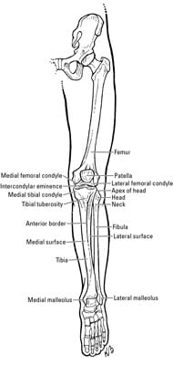
Clinical Anatomy The Bones Of The Knee And Leg Dummies from www.dummies.com This is the first joint in the leg. The forearm is the long bone that runs just after the elbow. The major bones of the leg are the femur (thigh bone), tibia (shin bone), and adjacent fibula, and these are all long bones.the patella (kneecap) is the sesamoid bone in front of the knee.most of the leg skeleton has bony prominences and margins that can be palpated and some serve as anatomical landmarks that define the extent of the leg. The tibia, commonly known as the 'shin bone', is the largest and most medial of the two.you can palpate its anterior border when you run your finger down the anterior aspect of your leg. Numerous bone is the long bone of the upper arm which goes all the way to the elbow. Long bone femur label 12 photos of the long bone femur label , bone. Some common causes of leg pain include: The bones of the leg are the femur, tibia, fibula and patella.
At the same time, the bones and joints of the leg and foot must be strong enough to support the body's weight while remaining.
The bones of the hip include the femur, the ilium, the ischium, and the pubis. The elbow is located below the chest at the back of the foreleg. At the same time, the bones and joints of the leg and foot must be strong enough to support the body's weight while remaining. Human anatomy diagrams show internal organs, cells, systems, conditions, symptoms and sickness information and/or tips for healthy living. The foot bones shown in this diagram are the talus, navicular, cuneiform, cuboid, metatarsals and calcaneus. The diagram of bones in the ankle and foot is given below: The bones of the leg are the femur, tibia, fibula and patella. The thigh bone, or femur, is the large upper leg bone that connects the lower leg bones (knee joint) to the pelvic bone (hip joint). 1 and the one behind the little toe is no. Premier leg skeleton with painted & labeled muscle attachments. The hip bone (os coxae, innominate bone, pelvic bone or coxal bone) is a large irregular bone, constricted in the center and expanded above and below leg bone diagram. It is made of the ulna and the radius. Some types of leg pain can be traced to problems in your lower spine.

